Chat with us, powered by LiveChat
 

Drummoyne Skin Cancer Centre

Our Géniale Skin Cancer doctors have over two decades of experience in medicine and surgery. Our centre is equipped with state-of-the-art technology and our team is highly skilled in the latest, most innovative skin cancer procedures.

SKIN EXAMINATION TYPES

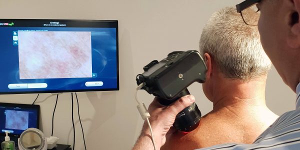
Géniale Skin Cancer Centre uses computerised dermoscopy and MoleMapping technology to evaluate, identify and monitor pigmented lesions in early stages, in order to distinguish malignant from benign skin lesions.

Our expert team invites you to make an appointment for a skin examination and to discuss the ideal skin cancer prevention and treatment for you with comprehensive screening, advanced diagnostics and technology for early diagnosis.

We’d Love To Hear From You!

Book your consult for early detection & treatment

Dermoscopy5

1. COMPUTERISED DERMOSCOPY

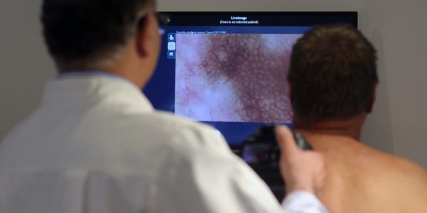
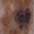
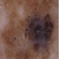
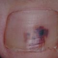
Computerised dermoscopy is a non-invasive diagnostic method used to examine pigmented skin lesions. It involves the use of high-resolution digital microscopy to allow our Géniale skin doctor to review and compare images taken at follow-up appointments.

Computerised dermoscopy enhances the accuracy of early detection of malignant skin cancers and reduces unnecessary biopsy or excision of benign skin lesions.

Dermoscopy

2. MOLEMAP – BODY PHOTOGRAPHY

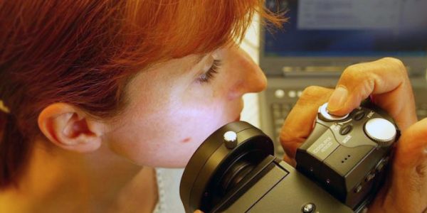
MoleMap imaging is a comprehensive skin check performed by a Géniale skin doctor. It is a procedure recommended and particularly useful for high-risk patients, patients with previous melanoma, or a large number of moles.

The MoleMap skin surface microscope creates a dermoscopic image and complete photographic record of your skin and all significant skin lesions. This enables us to monitor changes in your skin, including the early detection of melanoma.

Health Digital imaging

SPOT CHECK

Up to 50% of melanoma appears in new skin. If you are worried about a mole, it is important to get it checked. You may book an appointment for a Spot Check within 12 months of your MoleMap.

Monitoring changes over time

Géniale Skin Cancer Centre uses Sequential Digital Dermoscopic Imaging and Clinical Imaging to monitor lesions of concern through sequential review. Serial clinical imaging highlights changes to the internal and external structure of any pigmented skin lesions. Body Photography is used to identify suspicious lesions and allows for lesions of concern to be monitored; reducing unnecessary excisions as a result.

We’d Love To Hear From You!

Book online or call today with our highly trained doctors and advanced diagnostic technology.

TYPES OF SKIN CANCER

Skin cancer occurs when unrepaired DNA damage to skin cells (most often caused by ultraviolet radiation from sunshine or tanning beds) triggers mutations (genetic defects) that lead the skin cells to multiply rapidly and form malignant tumors.

Australia has the highest rate of skin cancer in the world. Two out of three Australians will be treated for skin cancer during their lives.

Too much UV exposure can cause irreversible damage to the DNA of skin cells and one damaged cell can start a deadly melanoma growing. Tanning, whether from sunlight or solariums can lead to skin damage, premature ageing, eye damage and skin cancers.

Skin cancers are grouped into two types, either non-melanoma or melanoma.  Non-melanoma skin cancers are the most common type of skin cancer and arise from the squamous and basal cells. These are referred to SCC and BCC respectively. A type of pre-cancerous growth of squamous cells is known as an actinic keratosis, or sunspot.

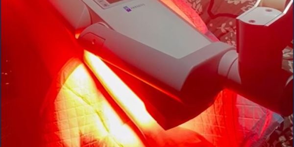
In cases where treatment is needed primarily for treating certain types of cancer and various skin conditions, Photodynamic Therapy (PDT) is an optional treatment choice. PDT is a non-surgical treatment for early cancerous lesions.

MELANOMA

MELANOMA

It is the most aggressive type of skin cancer and it is the only type of cancer with a death rate still on the rise.
BASAL CELL CARCINOMA (BCC)

BASAL CELL CARCINOMA (BCC)

It is the commonest type of skin cancer and represents 65% to 80% of skin cancer.
SQUAMOUS CELL CARCINOMA (SCC)

SQUAMOUS CELL CARCINOMA (SCC)

SCC is the second most common form of skin cancer.

1. MELANOMA

Early detection of this type of skin cancer can be difficult.  Although there are many different subtypes of melanoma clinically and histologically, they can ultimately be divided into two categories:

  • Fast growing melanoma, usually thick or nodular melanoma
  • Slow growing, usually thin or superficial spreading melanoma

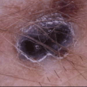
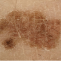
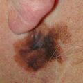
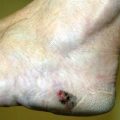
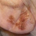
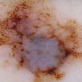
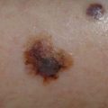
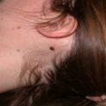
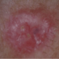
Examples of Melanoma:

Fast growing melanomas can be fatal within a few weeks.  In many instances, patients do not have a chance to present to their doctor before it is too late.  However, if it is detected and treated early enough there is a chance that it can be cured. Slow growing melanoma can take months or years to grow before it becomes fatal. It is very often indistinguishable from a normal mole with a naked eye therefore careful skin examination to detect this type of skin cancer is very important.  If you have regular skin checks no one should die from it.

2. BASAL CELL CARCINOMA (BCC)

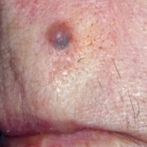
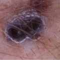
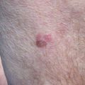
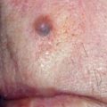
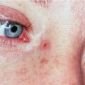
It is the commonest type of skin cancer and represents 65% to 80% of skin cancer. It occurs most commonly on the face and neck with raising incidence on the trunk. Appearances of basal cell carcinomas are often small round or flatten in shape and red, pale or pearly in colour. BCCs usually grow slowly but can become locally invasive and penetrate deeper tissue causing significant tissue destruction and disfigurement. Metastasis of this type of cancer is rare.

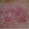
Examples of BCC:

3. SQUAMOUS CELL CARCINOMA (SCC)

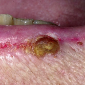
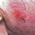
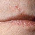
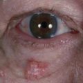
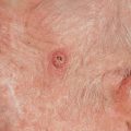
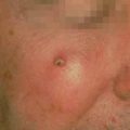
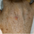
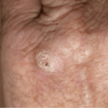
It usually arises in an area that has had some premalignant change such as solar keratosis secondary to sun damage. There is a strong correlation with damage to the skin by the sun and can be experimentally produced by ultraviolet light.  The appearances of these tumours are more inflammatory, indurated and ulcerate sooner compared with BCC.

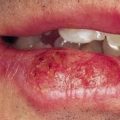
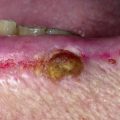
Examples of Squamous Cell Carcinoma (SCC):

Book Your Skin Cancer Check!

Get a complete record of your skin & monitor changes over time

"*" indicates required fields

Name*
This field is for validation purposes and should be left unchanged.

PREMALIGNANT CONDITIONS

1. ACTINIC KERATOSIS (AK) OR SOLAR KERATOSIS (SK)

It affects 40 – 60% of the Australian Caucasian population over 40 years of age and 80% of those aged between 60 and 69 years.  It is a premalignant condition often a precursor to squamous cell carcinoma (SCC).  Its appearances are usually discrete, erythematous, scaly or crusty patches of skin and for that reason are often indistinguishable to SCC. Therefore, anyone with multiple AK lesions should have regular follow up and be treated accordingly.

Actinic Keratosis can be difficult to distinguish from squamous cell carcinoma clinically. In most cases a punch biopsy or total lesion excisional biopsy is necessary to confirm a clinical diagnosis as the treatment options for these conditions will depend on it.

2. DYSPLATSTIC NAEVI & DYSPLASTIC NAEVUS SYNDROME

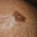
Dysplastic Naevi are high-risk moles and have a high potential to progress to malignant melanoma. The appearance of these moles are usually irregular, uneven distribution of colour, raised or flat, large moles which share some of the features of early melanoma. In the case of severely dysplastic naevi they should be treated as melanoma in situ and wider skin surgical excision is warranted.

Dysplastic Naevus Syndrome (DNS) is diagnosed when a person has 5 or more of dysplastic moles and they need to be confirmed histologically. Patients with DNS usually have multiple moles which make management more difficult. Therefore it is mandatory that everyone with DNS should be in a close monitoring program such as total body photography with digital monitoring and some of these moles should be removed and sent for histological confirmation to exclude melanoma.

The single most important determinant of whether an individual is prone to develop melanoma or not is the presence or absence of dysplastic naevi. There are numerous epidemiological studies that confirm the significance of dysplastic naevi as markers for increased risk for developing melanoma.

Book your skin check today

Category
Skin Cancer Centre

Book Your Obligation Free Consultation Today

Translate »